The defect in the skull base was in the medial medium part of the ethmoid roof.
Ethmoid roof defect.
Edges of skull base defect are identified and freshened.
Osteodural defects related to the sphenoid roof are rare.
The keros classification is a method of classifying the depth of the olfactory fossa.
Defects are encountered at the ethmoid roof cribriform plate and sphenoid sinus.
At times these defects can be missed during the course of the operation and this can be attributed by the defect size and csf fl ow rate.
Spontaneous traumatic or iatrogenic skull base defect.
The midline sphenoid sinus was the third most common location with 5 17 8 of 28 defects recognized in this location.
Fascia lata and adipose tissue grafts were taken to close the defect.
A skull base defect of the right ethmoid roof is evident with pulsations of the brain seen hanging into the right nasal cavity.
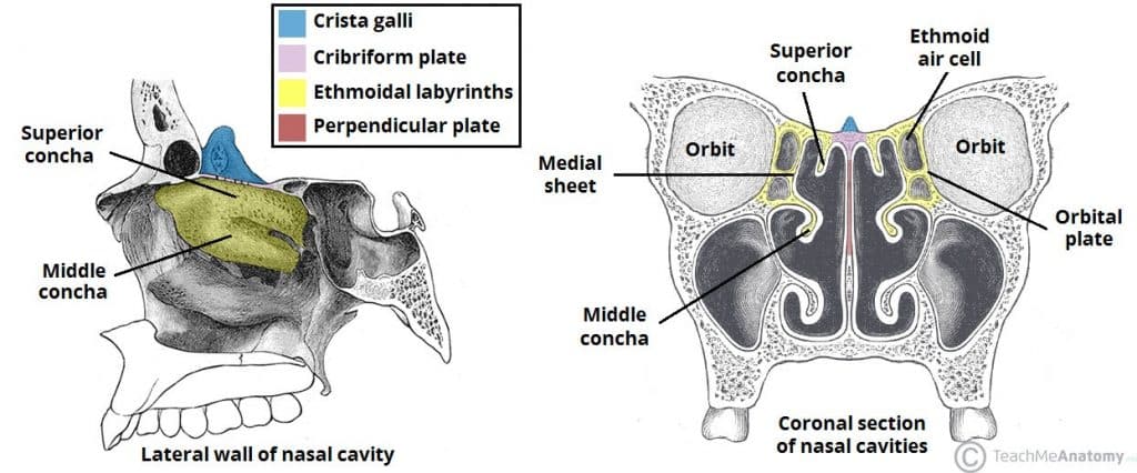
The ethmoid bone is one of the bones that make up the orbit of the eye.
References promoted articles advertising edit article share article.
The ethmoid bone forms the area of the skull at the roof of the nose where it sets the nasal cavity apart from the brain thus the ethmoid is right between the two orbits which contain the eyes.
The mucosal edges have been freshened and preparation is made for.
The size of the defect was the same as rope the diameter of nasal packing about 1 mm fig.
In our study in addition to the variations in ethmoidal roof and olfactory fossa we have analyzed the association of the olfactory fossa depth with the asymmetries of height and contour of the ethmoidal roof.
Asymmetry of the ethmoid roof is an important anatomic variation seen on ct scan that has the potential for disastrous complications in ess.
Underlay cartilage bone graft placed extra durally in large defects.
Report problem with article.
The roof was affected anterior to the tuberculum sellae in 3 of our patients and the posterior wall in 2 patients.
Accurate diagnosis is crucial to avoid the devastating consequences.
Graft is held firmly in place with absorbable packing material.
The very thin horizontal cribriform plate lamina cribrosa of the ethmoid bone is bounded laterally by the vertical lateral lamella.
The ethmoid labyrinth is covered by the fovea ethmoidalis of the frontal bone and separates the ethmoidal cells from the anterior cranial fossa.
The fovea ethmoidalis is an extension of the orbital plate of the frontal bone and forms the lateral part of the ethmoid roof.
From greek ethmos sieve is an unpaired bone in the skull that separates the nasal cavity from the brain it is located at the roof of the nose between the two orbits the cubical bone is lightweight due to a spongy construction.